Study Reveals Histone H3K9me2 & H3K79me3 as Potential Guardians Against Cancer
Epigenetic changes involving modifications to DNA and histones play a pivotal role in controlling gene expression,
shaping cellular identity, and governing cellular functions. These changes are central to processes like cell
differentiation and reprogramming, enabling cells to transition between different functional states. Among these
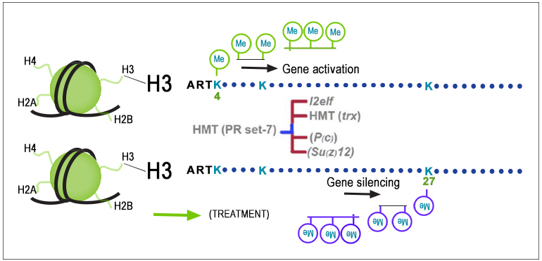
changes, histone modifications, particularly H3 methylation, are crucial regulators of gene activity. Some histone
marks, like H3K4me3 and H3K79me2/3, activate genes, while others, like H3K27me3 and H3K9me2/3, repress gene
activity. Enzymes known as histone-modifying enzymes (HMEs) add or remove these chemical marks on histones,
ultimately influencing the cell's gene expression profile.
These epigenetic changes are not only fundamental in normal cellular processes but also have important implications
in diseases like cancer. During the development of cancer (tumorigenesis), cells lose their distinctive
characteristics and acquire properties similar to stem cells. This transformation is accompanied by altered histone
modifications and DNA methylation patterns, highlighting a significant link between normal adult stem cells and
cancer.
In a recent study published in the International Journal of Molecular Sciences, researchers delved into the
H3 modifications occurring as neuroblastoma cells differentiate into osteoblasts (bone cells). They identified specific H3
methylation patterns critical to this process and observed differences in tissue samples from cancer patients
compared to healthy tissue. Their findings suggest that the simultaneous presence of high levels of H3K9me2 and
H3K79me3 could function as an epigenetic barrier against the development of cancer, offering a potential diagnostic
marker.
In a previous study, the researchers demonstrated that human neuroblastoma cells can transform into osteoblasts in just
five days. To understand the epigenetic changes underlying this process, they conducted a genome-wide analysis of
various histone modifications during differentiation, including different forms of histone H3 methylation, acetylation,
and phosphorylation. Using EpigenTek’s histone extraction kit (Cat # OP-0006), histone
multiplex assay (Cat # P-3100)
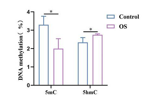
and DNA methylation ELISA assay (Cat # P-1030),
they uncovered a strong correlation between these specific H3 marks and changes in DNA methylation, thus highlighting
the role of chromatin remodeling in governing gene activity during cellular differentiation
Graphical representation of overlapping relationship between H3 methylation
changes (H3K4me1, H3K9me2, H3K27me3, H3K79me2) and global DNA methylation state, during
differentiation.[Piro MC et. al.
PMC10419041]
To expand the applicability of their findings to cancer, the researchers examined skin biopsy specimens from patients
with basal cell carcinoma (basalioma), a common type of skin cancer. Using immunofluorescence analysis with specific
antibodies, they compared the levels of distinct histone modifications, including H3K9me2 (Cat # A-4035),
H3K27me3 (Cat # A-4039),
H3K79me2 (Cat # A-4044),
and H3K79me3 (Cat # A-4045)
in both cancerous and healthy tissue regions. The striking differences in histone modification patterns suggest the
potential utility of these epigenetic marks as indicators of cancerous tissue.
b) Immunostaining of basalioma biopsies with H3K9me2, H3K27me3, H3K79me2, and
H3K79me3 antibodies. c) 4x magnification image of H3K9me2 antibody staining. d) Quantitative analysis of
positive nuclei for H3K9me2, H3K27me3, and H3K79me3 from immunofluorescence images. [Piro MC et. al.
PMC10419041]
Their investigation also encompassed other cancer types, such as head and neck tumors and urothelial bladder
carcinoma. This extensive analysis consistently uncovered noteworthy modifications in histone patterns associated
with cancer. Additionally, they identified robust correlations between specific histone marks, notably H3K9me2 and
H3K79me3, within cancerous tissues.
Bioinformatics was used to gain a deeper understanding of the biological mechanisms underlying differences in H3
marks. The research team scrutinized 14 enzymes responsible for modifying H3K9 and H3K79 histone marks in cancer
relative to normal tissues, with a focus on enzymes that write (methyltransferases), read, and erase (demethylases)
histone marks. This approach sheds light on potential regulatory pathways that contribute to the observed patterns
of histone modifications in cancer. It also supports the notion that elevated global levels of H3K9me2 and H3K79me3,
present in normal differentiated cells but absent in malignancy, may play a role in safeguarding against the
development of tumors.
Overall, the results from this study emphasize the significance of H3K9me2 and H3K79me3 as critical epigenetic
factors that help preserve normal cell identity and act as protective measures against cancer development. When
these epigenetic marks are downregulated in cancer tissues, it signifies a loss of cellular differentiation. The
study observed that H3K27me3 levels exhibited variations depending on the type of tumor, suggesting it may not
consistently serve as an epigenetic barrier. Nonetheless, the correlation between H3K9me2 and H3K79me3 underscores
their combined role in maintaining cellular differentiation. These epigenetic marks may serve as valuable diagnostic
indicators and potential targets for therapeutic interventions aimed at restoring a normal epigenome in cancer.


 Cart (0)