




Assisted reproductive technology (ART) has revolutionized fertility treatment, facilitating the conception of nearly 10 million children globally. However, concerns linger over its association with adverse gestational and perinatal outcomes, notably an increased incidence of low birth weight. Central to ART is ovarian stimulation (OS), a process that stimulates ovaries to produce multiple oocytes (superovulation), yet its implications for birth weight and placental function remain a subject of intense study.
Prior studies have linked low birth weight to placental deterioration, possibly stemming from disrupted genomic DNA methylation in oocytes caused by OS. However, the precise mechanisms by which OS leads to abnormal DNA methylation patterns in oocytes remain unclear.

In a study featured in the journal Cell Communication and Signaling, researchers have found a potential mechanism involved in the loss of DNA methylation in OS-induced oocytes. Their finding holds promise for enhancing safety and reducing epigenetic abnormalities in ART procedures.
DNA methylation, an essential epigenetic mechanism in embryonic development, involves attaching a methyl group to a cytosine preceding a guanine base (CpG). ART procedures can influence DNA methylation in oocytes, embryos, and fetuses. During oocyte growth, DNA methylation is dynamically regulated, and superovulation, a common aspect of ART, is thought to interfere with this process.
It has been suggested that OS may reduce maternal DNA methylation in oocytes during the methylation acquisition or maintenance processes. High estrogen levels during OS may also contribute to this loss of DNA methylation, affecting genes crucial for placental function.
In the current study, the researchers sought to investigate how OS affects DNA methylation in oocytes by exploring if changes are linked to abnormal development of the placenta, the organ that nourishes the developing baby. They hypothesized that OS might cause estrogen receptors (ERα) to activate the protein TET2, leading to DNA methylation changes.
Using mouse oocytes and mouse parthenogenetic embryonic stem cells, they assessed global levels of 5-methylcytosine (5mC) and 5-hydroxymethylcytosine (5hmC) using our highly specific 5mC and 5hmC monoclonal antibodies and MethylFlash™ Global DNA Methylation and Hydroxymethylation ELISA Easy Assays. Furthermore, they analyzed genome-wide methylation and the methylation status of the maternally imprinted Mest promoter region, a specific gene crucial for development that appears to be directly targeted by the ERα-TET2 complex.
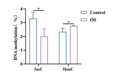
Their findings revealed that elevated estrogen levels resulting from OS led to a reduction in global 5mC levels and an increase in global 5hmC levels in oocytes. Similar changes were observed in early-stage fertilized oocytes (zygotes) and embryonic stem cells exposed to high estrogen. Remarkably, inhibiting estrogen signaling or eliminating TET2 prevented these methylation alterations, suggesting a potential role for TET2 in the process triggered by high estrogen.
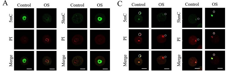
Moreover, the study uncovered evidence suggesting that TET2 may be directly influenced by estrogen receptors and may cooperate with them to demethylate specific genes under high estrogen conditions.
Overall, the study suggests a potential mechanism by which ovarian stimulation (OS) may disturb DNA methylation patterns in oocytes and early embryos. This disruption could impact gene expression, especially maternal imprinted genes, potentially leading to abnormal placental development and low birth weight. The findings provide important insights into the effects of fertility treatments on offspring health, paving the way for further investigation into the safety of ART procedures and the development of strategies to minimize risks associated with OS.
Data Image



