Choose the catalog number and quantity to pre-fill on the quote request page.
EpiQuik 8-OHdG DNA Damage Quantification Direct Kit (Colorimetric)
MethylFlash Hydroxymethylated DNA 5-hmC Quantification Kit (Colorimetric)
EpiQuik 8-OhG RNA Damage Quantification Direct Kit (Colorimetric)
Epigenase 5mC-Hydroxylase TET Activity/Inhibition Assay Kit (Colorimetric)
SOD1 Protein
KIR2DS1 Polyclonal Antibody
The EpiQuik™ 8-OHdG DNA Damage Quantification Direct Kit (Colorimetric) is a complete set of optimized buffers and reagents to colorimetrically detect and quantify oxidative DNA damage (8-OHdG) directly using DNA isolated from any species such as mammals, plants, fungi, bacteria, and viruses in a variety of forms including, but not limited to, cultured cells, fresh and frozen tissues, paraffin-embedded tissues, and body fluid samples. The kit has the following advantages and features:
Background Information8-hydroxy-2’-deoxyguanosine (8-OHdG or 8-oxo-dG) is an oxidized derivative of deoxyguanosine and is generated by hydroxyl radicals, singlet oxygen, and one-electron oxidants in cellular DNA. As a modified nucleoside base, 8-OHdG is considered important not only because of its abundance but also because of its mutagenic potential through G-to-T transversion mutations upon replication of DNA. 8-OHdG also participates in epigenetic regulation of gene activation/repression by inhibiting the binding affinity of MBD protein to the CpG sites of DNA. Currently, 8-OHdG is widely accepted as a sensitive marker of oxidative DNA damage and oxidative stress. Evidence shows that increased levels of 8-OHdG are closely correlated with exposure to harmful environmental factors such as ionizing radiation, industrial chemicals, air pollution, cigarette smoking, and cancer chemotherapy.
Several chromatography-based techniques such as HPLC-ED and LC-MS are used for detecting 8-OHdG in tissues and cells. However these methods are time consuming and have low throughput with high costs. The currently used competitive ELISA methods are also not conveniently applicable for cell/tissue 8-OHdG detection because they are less accurate and have an inability to use intact DNA isolated from cells or tissues directly.
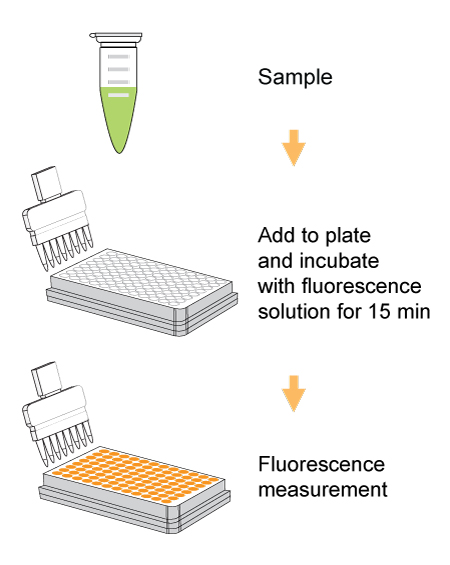
Principle & ProcedureThe EpiQuik™ 8-OHdG DNA Damage Quantification Direct Kit (Colorimetric) contains all reagents necessary for the quantification of Oxidative DNA damage (8-OHdG). In this assay, DNA is bound to strip wells that are specifically treated to have a high DNA affinity. 8-OHdG is detected using capture and detection antibodies. The detected signal is enhanced and then quantified colorimetrically by reading the absorbance in a microplate spectrophotometer. The amount of 8-OHdG is proportional to the OD intensity measured.
Starting Materials & Input AmountDNA amount can range from 100 ng to 300 ng per reaction. An optimal amount is 300 ng per reaction. Starting DNA may be in water or in a buffer such as TE.
Safe and ConvenientAll the necessary reagents, including negative controls and positive controls, for the quantification of 8-OHdG are conveniently packaged in the kit. The direct colorimetric quantification of DNA samples replaces obsolete or inferior methods and eliminates the need for DNA digestion/denaturation, radioactivity, extraction, or chromatography.
Responsive, Reliable, and PracticalBased on its working principle and the microplate format, the kit can be practically and routinely used for any species in a variety of forms including cultured cells, fresh and frozen tissues, and paraffin-embedded tissues. To demonstrate the capabilities of the kit, it has been successfully used for quantifying the content of 8-OHdG in DNA from human kidney, liver, and mouse brain tissues. The percentage of 8-OHdG measured by the kit is similar and comparable to that detected by HPLC methods (see Table 1 and Fig. 4).
2 ug completely digested DNA/assay


 Cart (0)