m6A RNA Methylation in Cancer Immunity and Therapeutic Resistance
Learn how m6A RNA methylation regulates tumor immunity, checkpoint response, and therapeutic resistance, and how to choose assays for global m6A quantification, MeRIP enrichment, and m6A-seq.
Therapeutic resistance is often studied through mutations, transcriptional programs, chromatin accessibility, DNA methylation, immune checkpoint expression, and tumor microenvironment remodeling. Those layers remain essential, but they do not fully explain how tumors rapidly adjust RNA stability, translation, splicing, immune signaling, stress adaptation, and antigen presentation. m6A RNA methylation, also known as N6-methyladenosine, adds a post-transcriptional regulatory layer that can shift how an existing transcriptome is used.
The practical question is no longer whether m6A is biologically important. Landmark m6A maps identified more than 12,000 m6A sites in transcripts from over 7,000 human genes, with enrichment near stop codons, long internal exons, and 3' UTRs. Another early m6A methylome study identified m6A-containing mRNAs from 7,676 mammalian genes and showed enrichment near stop codons and 3' UTRs, linking m6A to regions that influence RNA stability, localization, translation, and microRNA interaction [1,2].
For cancer biology, the stronger question is experimental: when a tumor becomes resistant to checkpoint blockade, chemotherapy, radiotherapy, or targeted therapy, does the resistant state involve global m6A remodeling, transcript-specific m6A redistribution, altered reader engagement, or a combination of these?
Because m6A can affect cancer immunity at multiple levels, from global RNA methylation changes to transcript-specific regulation of immune signaling, assay choice becomes part of the biological interpretation. A resistant tumor model with altered total m6A may require a different follow-up strategy than a study focused on one checkpoint pathway, one cytokine transcript, or one RNA-binding protein interaction. Before choosing a platform, researchers should define whether they need to measure overall m6A abundance, test candidate transcripts, map enriched regions across the transcriptome, or validate a specific mechanistic link between m6A and therapy response.
Choosing the right m6A method
m6A method selection should start with the question.
Use global m6A quantification when the question is whether total RNA methylation changes across biological states. This is appropriate for comparing untreated versus drug-treated cells, parental versus resistant cell lines, immune checkpoint-sensitive versus resistant models, or immune-stimulated versus unstimulated samples. A global assay is efficient for screening conditions, ranking samples, and deciding whether deeper mapping is justified. However, it does not identify which transcripts are modified or where m6A occurs.
Use m6A enrichment followed by qPCR when the question is focused on candidate transcripts. This approach is useful when prior RNA-seq, pathway analysis, literature evidence, or functional data point to specific immune or resistance-associated genes. Examples include transcripts involved in interferon signaling, antigen processing, cytokine response, lactate metabolism, apoptosis, DNA damage response, drug efflux, or stemness. Enrichment-qPCR can help determine whether candidate RNAs show altered m6A enrichment between sensitive and resistant states.
Use m6A enrichment followed by sequencing when the question is discovery-driven. This is the right choice when researchers need to identify m6A-enriched regions across the transcriptome, compare treatment-induced methylation landscapes, or discover pathways that were not predicted from gene expression alone. Sequencing-based approaches are more informative, but they also require stronger experimental design, including biological replicates, input controls, negative controls, RNA quality assessment, and careful bioinformatic analysis.
The core biology: writers, erasers, readers, and RNA fate
m6A is installed primarily by methyltransferase complexes that include METTL3 and METTL14, removed by demethylases such as FTO and ALKBH5, and interpreted by reader proteins such as YTH domain family proteins. Through those writer, eraser, and reader proteins, m6A can affect mRNA decay, translation, splicing, nuclear export, and RNA localization [3,4].
That versatility is why m6A can generate apparently different outcomes in different cancers. A reader that promotes decay of one transcript may reduce an immune stimulatory pathway in one context but suppress an oncogenic pathway in another. A writer such as METTL3 can support tumor growth in some models while also supporting immune cell function in others. In cancer immunity and resistance research, it is therefore risky to treat "more m6A" or "less m6A" as a simple directional biomarker. The more useful approach is to define which cell compartment, transcript class, and treatment condition is being studied.
m6A and tumor antigen presentation
One of the clearest links between m6A and cancer immunity comes from dendritic cell antigen presentation. In a Nature study, YTHDF1 was shown to suppress anti-tumor immunity by enhancing translation of lysosomal proteases in dendritic cells. These proteases can limit cross-presentation of tumor antigens. Loss of YTHDF1 improved antigen-specific CD8+ T cell responses and enhanced the therapeutic effect of PD-L1 checkpoint blockade in mouse models [5].
This finding illustrates why m6A studies in immuno-oncology should not be limited to tumor cells. The same tumor sample contains cancer cells, dendritic cells, macrophages, T cells, NK cells, stromal cells, and endothelial cells. Bulk RNA can reveal a treatment-associated change, but interpreting that change requires attention to cellular composition. When possible, researchers should pair m6A data with immune cell profiling, sorted populations, single-cell data, or orthogonal markers of antigen presentation.
For discovery projects, the EpiQuik CUT&RUN m6A RNA Enrichment (MeRIP) Kit (#P-9018) can support a focused MeRIP-style enrichment strategy when researchers already have candidate genes or need enriched RNA for downstream PCR or sequencing. Its CUT&RUN-based enrichment workflow is designed for low input RNA and can enrich m6A-containing fragments for region-specific PCR or epitranscriptome-wide profiling.
m6A in T cells, Tregs, NK cells, and immune pressure
m6A biology also affects immune effector and suppressor cell function. In T cells, METTL3-mediated m6A has been linked to IL-7/STAT5/SOCS pathway regulation and T cell homeostasis [6]. In regulatory T cells, m6A methylation helps sustain suppressive function through pathways involving SOCS transcripts and IL-2/STAT5 signaling [7]. In NK cells, METTL3-mediated m6A promotes anti-tumor immunity by supporting NK cell response to IL-15; Mettl3 deletion impaired NK cell infiltration and function in the tumor microenvironment and accelerated tumor development in mice [8].
These examples explain why m6A can influence both immune activation and immune suppression. In one immune compartment, m6A loss may impair effector function. In another, it may weaken suppressive activity. A strong experimental design should therefore define whether the primary question is tumor-intrinsic resistance, immune cell dysfunction, or the interaction between both.
m6A and checkpoint resistance
Several studies now connect m6A regulators to response or resistance to immune checkpoint blockade. FTO has been reported to promote melanoma tumorigenicity and resistance to anti-PD-1 blockade. The study linked FTO activity to reduced m6A levels on target mRNAs and suggested that combining FTO inhibition with anti-PD-1 blockade may reduce resistance in melanoma models [9].
ALKBH5 has also been implicated in anti-PD-1 response. A PNAS study reported that ALKBH5 affects m6A density and splicing events during immune checkpoint blockade, modulates Mct4/Slc16a3 expression and lactate in the tumor microenvironment, and influences suppressive Treg and myeloid-derived suppressor cell accumulation [10].
In another study, depletion of the m6A methyltransferases Mettl3 and Mettl14 enhanced response to anti-PD-1 therapy in colorectal carcinoma and melanoma models, with the work highlighting IFN-gamma-Stat1-Irf1 signaling and Ythdf2-associated transcript regulation. The same publication noted that mismatch-repair-proficient or microsatellite instability-low colorectal cancers (CRC) represent about 85% of CRC patients, underscoring the interest in mechanisms that may help explain poor checkpoint response in low mutation burden settings [11].
The key takeaway for researchers is that m6A regulation can sit between tumor stress biology and immune recognition. It may influence cytokine signaling, antigen processing, suppressive cell recruitment, metabolic adaptation, and checkpoint sensitivity.
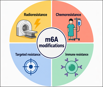
m6A and broader therapeutic resistance
m6A is not limited to immunotherapy resistance. Reviews of m6A in cancer therapy resistance describe roles in chemotherapy response, radiotherapy resistance, targeted therapy resistance, cancer stem-like states, epithelial-to-mesenchymal transition, apoptosis, DNA damage response, and drug transporter regulation [12,13].
This matters for combination therapy studies. A tumor that survives chemotherapy may later show altered immune recognition. A tumor exposed to checkpoint pressure may adapt through stress pathways that also affect drug sensitivity. Because m6A can influence RNA fate rapidly, measuring m6A before and after treatment can help distinguish stable genomic resistance from more plastic RNA-level adaptation.
A practical starting point is to use global m6A quantification to determine whether resistant and sensitive models differ in overall m6A level. The EpiQuik m6A RNA Methylation Quantification Kit (#P-9005) provides a colorimetric format for direct m6A RNA methylation quantification using total RNA. It includes negative and positive RNA controls and allows absolute or relative m6A RNA methylation assessment. For labs needing fluorescence readout, the EpiQuik m6A RNA Methylation Quantification Kit (Fluorometric) (#P-9008) offers a more sensitive fluorometric version.
Global assays do not identify modified transcripts or modification sites. They are best used for screening treatment conditions, comparing matched sensitive and resistant models, or prioritizing samples for deeper enrichment or sequencing.
Experimental design recommendations
For cancer immunity and resistance studies, m6A experiments are strongest when they include matched biological states. Examples include parental versus resistant cells, untreated versus drug-treated cells, immune checkpoint-sensitive versus resistant models, normoxia versus hypoxia, nutrient-replete versus nutrient-stressed cells, or co-culture conditions with immune cells.
RNA quality is critical. Degraded RNA can distort enrichment, reduce library quality, and complicate interpretation. DNA contamination should be minimized, especially when downstream readouts include qPCR or sequencing. When comparing global m6A levels, use equal RNA input and replicate samples. When performing enrichment, include input RNA, IgG or non-immune controls, and positive controls where appropriate.
A useful study sequence is:
Screen total RNA m6A levels with P-9005 or P-9008 across sensitive and resistant states.
Use P-9018 to test candidate immune or resistance transcripts by enriched RNA-qPCR.
Move to P-9016 when broad transcriptome-wide discovery is needed.
Validate candidate m6A changes with orthogonal RNA expression, protein, pathway, and functional assays.
This staged workflow reduces the chance of overinterpreting a single assay. A global m6A increase may be biologically important, but it does not reveal which transcripts changed. A MeRIP peak can indicate enrichment, but it should be interpreted with input RNA, expression level, antibody specificity, fragment size, and biological replicate consistency in mind. Published work has highlighted reproducibility and analysis limits in MeRIP/m6A-seq, making careful controls and validation especially important [14].
Product Selector for m6A Cancer Immunity and Resistance Studies
Research Need
Relevant Product
Best Fit in the Workflow
Key Notes
Screen global m6A changes across treatment, immune stimulation, or resistant models
Designed for region-specific m6A analysis by PCR or epitranscriptome-wide profiling by NGS; input range 1 ug to 20 ug, data obtainable from as low as 500 ng total RNA.
Transcriptome-wide discovery with integrated library preparation
Species-independent rabbit monoclonal antibody, clone 2H6; applications include ELISA, DB, IF, MeRIP, and nucleotide array.
m6A RNA methylation gives cancer researchers a practical way to study how tumor cells and immune cells adjust RNA behavior under therapeutic pressure. The field is moving from descriptive catalogs of m6A sites toward functional questions: which m6A-regulated transcripts shape antigen presentation, immune suppression, interferon signaling, metabolic adaptation, stemness, or drug response?
The most useful studies will not rely on a single m6A readout. They will combine global quantification, transcript-specific enrichment, sequencing, RNA expression, protein-level validation, and perturbation of writers, erasers, or readers. For cancer immunity and therapeutic resistance, that integrated approach can help clarify whether m6A is a marker of treatment response, a mechanistic driver of resistance, or a context-dependent vulnerability worth deeper investigation.
References
Dominissini D, et al. Topology of the human and mouse m6A RNA methylomes revealed by m6A-seq. Nature. 2012. View article
Meyer KD, et al. Comprehensive analysis of mRNA methylation reveals enrichment in 3' UTRs and near stop codons. Cell. 2012. View article
Song H, et al. METTL3-mediated m6A RNA methylation promotes the anti-tumour immunity of natural killer cells. Nature Communications. 2021. View article
Yang S, et al. m6A mRNA demethylase FTO regulates melanoma tumorigenicity and response to anti-PD-1 blockade. Nature Communications. 2019. View article
Li N, et al. ALKBH5 regulates anti-PD-1 therapy response by modulating lactate and suppressive immune cell accumulation in tumor microenvironment. PNAS. 2020. View article
Wang L, et al. m6A RNA methyltransferases METTL3/14 regulate immune responses to anti-PD-1 therapy. EMBO Journal. 2020. View article
Wang D, et al. N6-methyladenosine (m6A) in cancer therapeutic resistance: Potential mechanisms and clinical implications. Biomedicine & Pharmacotherapy. 2023. View article
Uddin MB, et al. Epitranscriptomic RNA m6A modification in cancer therapy resistance: Challenges and unrealized opportunities. Advanced Science. 2025. View article
McIntyre ABR, et al. Limits in the detection of m6A changes using MeRIP/m6A-seq. Scientific Reports. 2020. View article


 Cart (0)