Choose the catalog number and quantity to pre-fill on the quote request page.
MethylFlash 5-Formylcytosine (5-fC) DNA Quantification Kit (Colorimetric)
MethylFlash Methylated DNA 5-mC Quantification Kit (Colorimetric)
MethylFlash Hydroxymethylated DNA 5-hmC Quantification Kit (Colorimetric)
Omega-Gliadin Polyclonal Antibody, FITC Conjugated
Histone H4K20me2 Dimethyl Peptide
Histone H3K79me2 (H3K79 Dimethyl) Monoclonal Antibody [Q7]
The MethylFlash™ 5-Formylcytosine (5-fC) DNA Quantification Kit (Colorimetric) is a complete set of optimized buffers and reagents to colorimetrically quantify 5-formylcytosine (5-fC) in a microplate-based format. It is suitable for use with DNA isolated from any species (mammals, plants, fungi, bacteria, viruses, etc.) in a variety of forms including, but not limited to, cultured cells, fresh and frozen tissues, paraffin-embedded tissues, and body fluid samples. The kit has the following advantages and features:
Background Information5-formylcytosine (5-fC) has recently been found in mammalian tissue and cells and has been classified as a seventh DNA base. It is formed by the oxidation of 5-hydroxymethylcytosine through TET hydroxylases and has been demonstrated to be an intermediate of the DNA demethylation process. The function of 5-fC in gene regulation is not yet entirely clear, but it has been shown that 5-fC exhibits replication-dependent dilution during mouse pre-implantation development and could be functional in regulating pre-implantation development as a whole. The detection of 5-fC in various tissues and cells is important because 5-fC could be a marker to indicate active DNA demethylation. 5-fC can also be directly excised by Thymine DNA glycosylase (TDG) to allow subsequent base excision repair (BER) processing which converts modified cytosine back to its unmodified state.
Several chromatography-based techniques such as HPLC-MS are used for the detection of 5-fC in tissue and cells. However, these methods are time-consuming, require large amounts of DNA and have low throughput with high costs. To address these problems, Epigentek offers the MethylFlash™ 5-Formylcytosine (5-fC) DNA Quantification Kit (Colorimetric), which uses a unique microplate-based procedure to directly quantify 5-fC.
Principle & ProcedureThe MethylFlash™ 5-Formylcytosine (5-fC) DNA Quantification Kit (Colorimetric) contains all the reagents necessary for the quantification of 5-fC. In this assay, DNA is bound to strip-wells that are specifically treated to have a high DNA affinity. 5-fC is detected using capture and detection antibodies. The detected signal is enhanced and then quantified colorimetrically by reading the absorbance in a microplate spectrophotometer at a wavelength of 450 nm. The amount of 5-fC is proportional to the OD intensity measured.
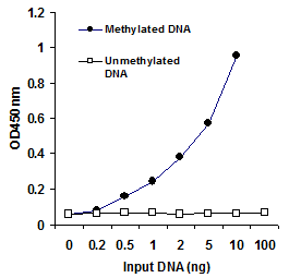
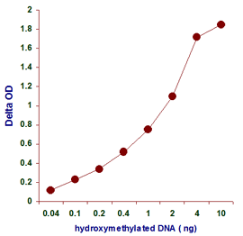
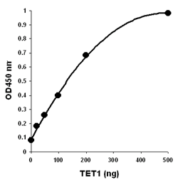
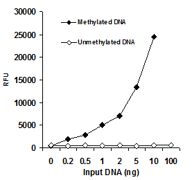
Starting Materials & Input AmountThe detection limit of the input DNA can be as low as 1 pg of 5-fC with a dynamic range from 10 pg to 400 pg (see Fig. 2). There is no cross-reactivity to cytosine, 5-mC, or 5-hmC within the indicated concentration range of the sample DNA (see Fig. 3).
Safe and ConvenientAll the necessary reagents, including negative and positive controls, for the quantification of 5-fC are conveniently packaged in the kit. The direct colorimetric quantification of DNA samples in a 96-well microplate format eliminates the need for DNA digestion/denaturation, radioactivity, extraction, or chromatography.
Easy, Fast, and FlexibleThe entire colorimetric assay has easy-to-follow steps for convenience and speed, allowing it to be completed in just 3 hours and 45 minutes. The strip-well microplate format allows for a flexible assay for manual or high throughput analysis. Universal positive and negative controls are included with the kit for the quantification of 5-fC from any species, such as mammals, plants, fungi, bacteria, and viruses.
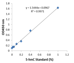
Responsive, Reliable, and PracticalBased on its working principle and the microplate format, the kit can be practically and routinely used for any species and for a variety of forms including cultured cells, fresh and frozen tissues, and paraffin-embedded tissues. To demonstrate the capabilities of the kit, it has been successfully used for quantifying the content of 5-fC in DNA from human brain tissue, mouse brain tissue, rectal tissue, and rectal cancer. The percentage of 5-fC measured by the kit is parallel and comparable to that detected by LC-MS/MS methods (see Fig. 4).


 Cart (0)