 Our single B-cell derived recombinant rabbit monoclonal antibodies have vastly improved reliability and reproducibility.



 Cart (0)


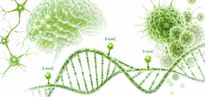
A combined methylation signal can conceal active remodeling
A conventional bisulfite result can be technically correct while collapsing two chemically and biologically different cytosine states into one value. Bisulfite treatment preserves both 5-methylcytosine, or 5-mC, and 5-hydroxymethylcytosine, or 5-hmC, as cytosine when read. Unless the method includes an additional step that separates them, the reported signal represents modified cytosine rather than 5-mC alone.
That ambiguity is consequential in brain tissue, differentiating cells, and cancer. A cytosine can shift from 5-mC to 5-hmC while remaining visible as a protected cytosine in standard bisulfite data. The combined signal may appear unchanged even though TET-dependent oxidation is occurring.
5-hmC entered mammalian epigenetics research in 2009, when it was detected at substantial levels in Purkinje neurons and other brain regions [1]. In parallel, Tahiliani and colleagues identified TET1 as an Fe(II)- and 2-oxoglutarate-dependent enzyme that oxidizes 5-mC to 5-hmC [2].
Further TET-dependent oxidation produces 5-formylcytosine and 5-carboxylcytosine, which can participate in pathways that restore unmodified cytosine. 5-hmC is therefore connected to DNA demethylation, but it does not function only as a short-lived reaction intermediate. Its abundance and genomic distribution can persist in stable, tissue-specific patterns.
A whole-genome analysis of 19 human tissue types found that 5-hmC was enriched in gene bodies and reflected gene expression more closely than gene-body 5-mC. Approximately one-third of the detected 5-hmC peaks were classified as tissue-specific differentially hydroxymethylated regions. Many were located near regulatory elements associated with tissue-specific gene programs [3].
These findings support a more precise experimental vocabulary. Global 5-hmC abundance describes how much of the mark is present. Hydroxymethylome profiling describes where it is distributed. Neither measurement substitutes for the other.
Neuronal genomes carry dense, structured 5-hmC
Brain contains substantially more 5-hmC than most other mammalian tissues. The modification is particularly prominent in postmitotic neurons, where DNA methylation patterns continue to change during maturation and functional specialization.
High abundance alone does not explain its research value. Neuronal 5-hmC is organized across genes and regulatory regions rather than dispersed uniformly through the genome. Its distribution varies among neuronal populations, developmental stages, and brain regions.
Single-cell methods have exposed differences that bulk methylation measurements cannot resolve. Joint single-nucleus hydroxymethylcytosine sequencing, or Joint-snhmC-seq, measures 5-hmC and true 5-mC separately in the same single-nucleus workflow. Applied to mouse brain, the method identified extensive heterogeneity across neuronal and non-neuronal populations. Separate 5-hmC and 5-mC profiles improved the identification of neuronal subtypes and revealed modification-specific relationships with cell-type-associated genes [4].
SIMPLE-seq provides another route to joint 5-mC and 5-hmC analysis at single-cell and single-molecule resolution. Its application to mouse brain, embryonic stem cells, and human peripheral blood mononuclear cells identified combinations of the two marks associated with different cell types and regulatory states [5].
The findings help explain why a combined methylation value may be uninformative in neuronal studies. Two neuronal populations can carry similar total levels of modified cytosine while differing in the relative contribution and genomic placement of 5-mC and 5-hmC.
Bulk brain studies face an additional problem. A tissue sample contains neurons, astrocytes, oligodendrocytes, microglia, vascular cells, and other populations. Differences between experimental groups may arise from altered hydroxymethylation within a cell type, a change in cellular composition, or both.
A 2025 study profiled 5-hmC across 1,079 autopsied dorsolateral prefrontal cortex samples. From 197,765 identified 5-hmC regions, the researchers reported 2,821 differentially hydroxymethylated regions associated with Alzheimer's disease neuropathology after adjustment for covariates and multiple testing. Several regions overlapped known Alzheimer's disease loci [6].
Those results describe associations in postmortem brain, not a diagnostic test. They also illustrate the difference between a regional hydroxymethylation map and a global measurement. Total 5-hmC can establish whether a broad shift has occurred. Regional profiling is needed to identify the genes or regulatory elements involved.
Tumor 5-hmC changes are global and regional
Many tumors contain less 5-hmC than their corresponding normal tissues. Reduced TET expression or activity, changes in cellular metabolism, altered availability of TET cofactors, and rapid proliferation can all contribute to this loss.
Melanoma supplied an early example. Lian and colleagues found extensive depletion of the normal 5-hmC landscape in melanoma and linked the change to disruption of the IDH-TET pathway. Restoration experiments in model systems partially rebuilt the 5-hmC landscape and reduced malignant properties [7].
A low global value does not mean that all genomic regions lose 5-hmC at the same rate. Tumor-associated hydroxymethylation can be redistributed while overall abundance declines.
Xue and colleagues examined tumor and normal tissues together with cell-free DNA across breast, colon, lung, ovarian, and pancreatic cancers. The study included 217 tumor tissues, 50 normal tissues, cell-free DNA from 1,009 participants with cancer, and 1,678 non-cancer participants. Global 5-hmC abundance decreased across the tumor types, while early-stage tumors already showed extensive regional redistribution. Some differentially hydroxymethylated regions were shared across cancers; others retained tissue-specific patterns [8].
A global assay and a genome-wide enrichment study would therefore describe different features of the same tumor. One records the overall loss of the mark. The other identifies the regions that lose, retain, or gain hydroxymethylation.
Separate measurement of 5-mC and 5-hmC can also reveal directional changes during tumor evolution. A 2026 colorectal cancer study analyzed cell-free DNA from 37 treatment-naive patients and 32 healthy controls using six-base whole-genome sequencing. In this exploratory cohort, a classifier combining 5-mC and 5-hmC achieved an area under the curve of 0.95, compared with 0.66 for a model that treated them as a combined modified-cytosine signal. Almost half of the examined differentially methylated regions showed increased 5-hmC in stage I disease and decreased 5-mC in stage IV disease [9].
The cohort was small and the findings require independent validation. The mechanistic observation is still relevant to experimental planning: a combined value can erase a transition between two cytosine modifications.
Bisulfite data require precise labeling
Standard bisulfite sequencing converts unmodified cytosine to uracil while preserving both 5-mC and 5-hmC as cytosine. WGBS, RRBS, targeted bisulfite sequencing, and bisulfite PCR inherit this chemical limitation unless another treatment is added.
Results from these methods are often described as DNA methylation data. In a 5-hmC-rich sample, "modified cytosine" or "combined 5-mC and 5-hmC" is more accurate.
Oxidative bisulfite sequencing addresses the ambiguity by oxidizing 5-hmC before bisulfite conversion. Comparison of oxidative bisulfite and conventional bisulfite data permits separate estimation of 5-mC and 5-hmC at single-base resolution [10]. Enzymatic and direct-detection methods now provide additional routes to modification-resolved sequencing.
High-resolution sequencing is warranted when the hypothesis concerns individual cytosines, enhancers, allele-specific patterns, or direct 5-mC-to-5-hmC remodeling. Many studies need a broader answer first: whether total 5-hmC differs between sample groups.
Matching the measurement to the hypothesis
Total genomic 5-hmC
Global quantification is suited to comparisons among treatments, tissues, cell states, disease models, or differentiation stages. The result describes the hydroxymethylated fraction of the DNA sample without identifying the contributing loci.
The MethylFlash Global DNA Hydroxymethylation (5-hmC) ELISA Easy Kit (P-1032) provides a colorimetric measurement of global 5-hmC in purified DNA. It can support initial screening, sample-group comparisons, and follow-up studies in which overall hydroxymethylation is the defined endpoint. A P-1032 result should not be used to assign hydroxymethylation to a specific gene or genomic region.
Hydroxymethylated regions
Regional studies require enrichment or sequencing. Hydroxymethylated DNA immunoprecipitation enriches DNA fragments that contain 5-hmC, after which candidate loci can be examined by PCR or other compatible downstream methods.
The EpiQuik Hydroxymethylated DNA Immunoprecipitation (hMeDIP) Kit (P-1038) supports fragment-level 5-hmC enrichment. It fits experiments in which the candidate genes or genomic regions have already been selected. The output reflects enrichment across DNA fragments rather than the modification percentage of an individual cytosine.
Cell and tissue distribution
Immunofluorescence and immunohistochemistry retain spatial information that is lost after bulk DNA extraction. These approaches can show whether a 5-hmC signal is concentrated in particular cells, tissue compartments, or morphological regions.
The 5-Hydroxymethylcytosine (5-hmC) Monoclonal Antibody [HMC/4D9] (A-1018) supports immunofluorescence, immunohistochemistry, dot blot, ELISA, and hMeDIP applications. Experimental controls remain essential because staining intensity and accessibility are influenced by sample preparation and assay conditions.
Parallel global 5-mC and 5-hmC
Separate global measurements can test whether the balance between the two marks changes across sample groups. This can be useful in differentiation studies, TET perturbation experiments, and tumor models where a shift in cytosine modification state is expected.
P-1032 can be paired with the MethylFlash Global DNA Methylation (5-mC) ELISA Easy Kit, P-1030, to obtain separate global 5-hmC and 5-mC readouts. The comparison does not prove that 5-mC was converted to 5-hmC at the same genomic sites. That conclusion requires locus-resolved or base-resolved analysis.
5-hmC workflow selector
| Research question | Measurement | Relevant kit | Interpretive boundary |
|---|---|---|---|
| Does total 5-hmC differ among sample groups? | Global 5-hmC quantification | P-1032 | Reports overall abundance, not genomic position |
| Are candidate regions associated with 5-hmC? | hMeDIP followed by downstream analysis | P-1038 | Enriches modified fragments, not individual bases |
| Which cells or tissue regions contain 5-hmC? | Antibody-based imaging or detection | A-1018 | Preserves spatial context but does not produce a genome-wide map |
| Do global 5-mC and 5-hmC change differently? | Separate global measurements | P-1032 and P-1030 | Shows sample-level balance, not direct locus-specific conversion |
| Which cytosines carry 5-mC or 5-hmC? | Modification-resolved sequencing | Specialized sequencing method | Provides base-level information with greater workflow and analysis demands |
Interpretation checks before drawing a biological conclusion
Cell composition can dominate a bulk result. A lower 5-hmC value in diseased brain tissue may reflect loss of a 5-hmC-rich neuronal population, a change within surviving neurons, or both. Cell sorting, single-cell analysis, histology, or cell-type marker data can help separate these possibilities.
An unchanged global value does not exclude regional remodeling. Gains and losses at different loci can offset one another when summed across the genome.
hMeDIP reports relative enrichment of 5-hmC-containing fragments. Enrichment is influenced by modification density, fragment size, antibody recognition, and background recovery. It should not be presented as a direct percentage of hydroxymethylation at a single cytosine.
Parallel global 5-mC and 5-hmC measurements provide two independent sample-level values. A reciprocal change is consistent with altered methylation and hydroxymethylation balance, but it does not establish a precursor-product relationship at defined loci.
5-hmC belongs in the assay decision, not only in the discussion
Neurobiology and cancer research frequently involve systems in which cytosine modification is being remodeled. Treating all protected cytosines as 5-mC can conceal that activity.
Global quantification, regional enrichment, spatial detection, and base-resolved sequencing occupy different positions in the workflow. The right choice depends on whether the study needs abundance, location, cellular context, or chemical identity.
A clear definition of the intended readout prevents a common interpretive error: asking a global or combined-modification method to support a locus-specific or 5-mC-specific conclusion.
References
- Kriaucionis S, Heintz N. The nuclear DNA base 5-hydroxymethylcytosine is present in Purkinje neurons and the brain. Science. 2009;324(5929):929-930. doi:10.1126/science.1169786. View article
- Tahiliani M, Koh KP, Shen Y, et al. Conversion of 5-methylcytosine to 5-hydroxymethylcytosine in mammalian DNA by MLL partner TET1. Science. 2009;324(5929):930-935. doi:10.1126/science.1170116. View article
- He B, Zhang C, Zhang X, et al. Tissue-specific 5-hydroxymethylcytosine landscape of the human genome. Nat Commun. 2021;12(1):4249. Published 2021 Jul 12. doi:10.1038/s41467-021-24425-w. View article
- Fabyanic EB, Hu P, Qiu Q, et al. Joint single-cell profiling resolves 5mC and 5hmC and reveals their distinct gene regulatory effects. Nat Biotechnol. 2024;42(6):960-974. doi:10.1038/s41587-023-01909-2. View article
- Bai D, Zhang X, Xiang H, Guo Z, Zhu C, Yi C. Simultaneous single-cell analysis of 5mC and 5hmC with SIMPLE-seq. Nat Biotechnol. 2025;43(1):85-96. doi:10.1038/s41587-024-02148-9. View article
- Zhao J, Gu T, Gao C, et al. Brain 5-hydroxymethylcytosine alterations are associated with Alzheimer's disease neuropathology. Nat Commun. 2025;16(1):2842. Published 2025 Mar 22. doi:10.1038/s41467-025-58159-w. View article
- Lian CG, Xu Y, Ceol C, et al. Loss of 5-hydroxymethylcytosine is an epigenetic hallmark of melanoma. Cell. 2012;150(6):1135-1146. doi:10.1016/j.cell.2012.07.033. View article
- Xue Y, Ning Y, Friedl V, et al. 5-hydroxymethylcytosine analysis reveals stable epigenomic changes in tumor tissue that enable cancer detection in cell-free DNA. Commun Biol. 2025;8(1):1613. Published 2025 Nov 19. doi:10.1038/s42003-025-09017-4. View article
- Puddu F, Johansson A, Modat A, et al. 5-methylcytosine and 5-hydroxymethylcytosine are synergistic biomarkers for early detection of colorectal cancer. Commun Med (Lond). 2026;6(1):15. Published 2026 Jan 20. doi:10.1038/s43856-025-01278-8. View article
- Booth MJ, Branco MR, Ficz G, et al. Quantitative sequencing of 5-methylcytosine and 5-hydroxymethylcytosine at single-base resolution. Science. 2012;336(6083):934-937. doi:10.1126/science.1220671. View article



