




A DNA methylation profile describes the modified cytosines present when a sample is collected. It does not reveal the full sequence of events that produced that profile.
Lower 5-methylcytosine, or 5-mC, may follow reduced DNA methyltransferase activity, incomplete maintenance after replication, increased TET-mediated oxidation, or expansion of a less-methylated cell population. Lower 5-hydroxymethylcytosine, or 5-hmC, may reflect weaker TET activity, limited 5-mC substrate, rapid cell division, continued oxidation, or changes in tissue composition.
Mechanistic studies can separate these possibilities by examining catalytic activity, global modified-base abundance, and genomic location in matched samples. The first measurement tests enzyme function. The second shows whether the total 5-mC or 5-hmC pool has changed. The third identifies the promoters, enhancers, genes, or CpGs involved.
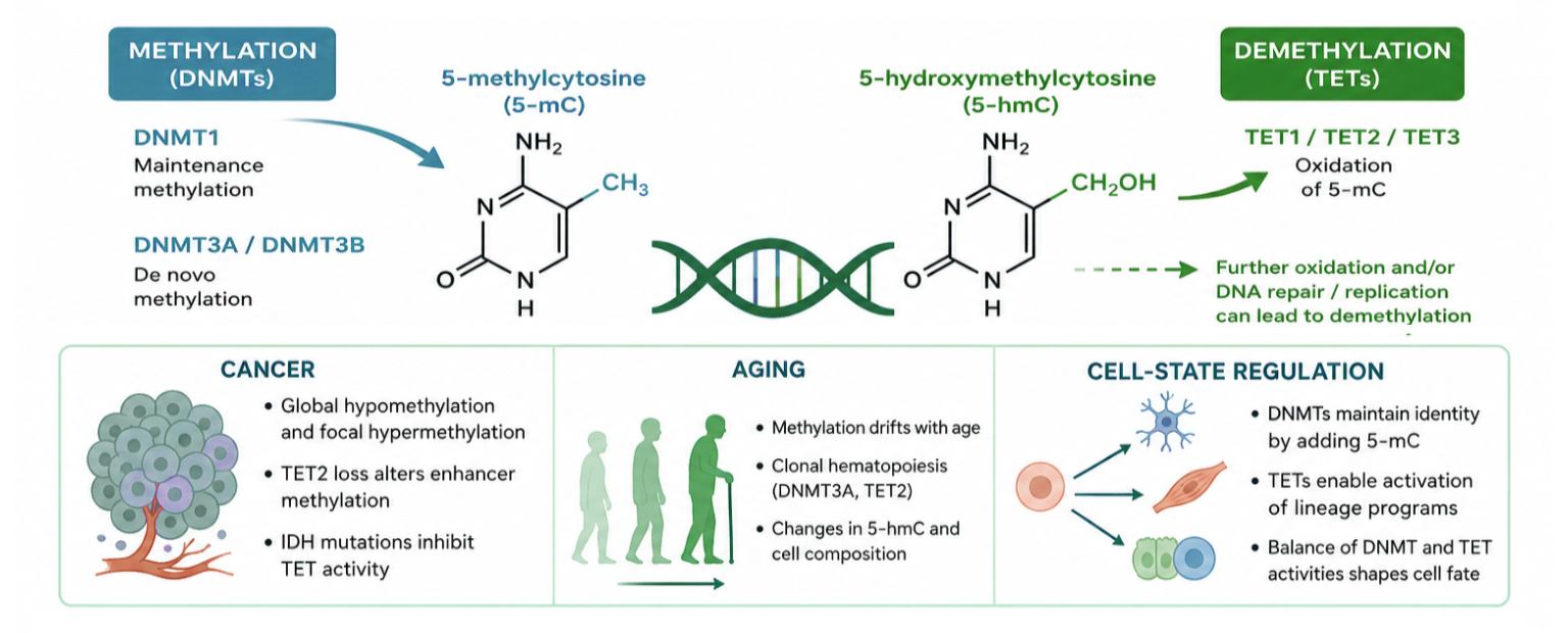
DNMT and TET Enzymes Define Methylation Turnover
DNMT1 preferentially restores methylation after DNA replication. DNMT3A and DNMT3B establish de novo methylation patterns and also contribute to maintenance at selected genomic regions. Histone modifications, transcription factors, chromatin accessibility, replication timing, and protein interactions influence where each enzyme acts.
TET1, TET2, and TET3 oxidize 5-mC to 5-hmC. Additional oxidation can produce 5-formylcytosine and 5-carboxylcytosine. These oxidized bases may be removed through DNA repair or diluted during replication, allowing methylated cytosines to return to an unmodified state.
Modified-base abundance may remain stable even when methylation is being actively added and removed. The iDEMS method, which combines metabolic labeling with quantitative mass spectrometry, showed that methylation maintenance can lag behind replication in mouse embryonic stem cells. Hydroxymethylation remained strongly biased toward parental DNA strands rather than being distributed equally across newly replicated sister strands [1].
The EpiQuik DNMT Activity/Inhibition ELISA Easy Kit (P-3139) can be used to compare total DNMT activity across matched nuclear extracts or purified enzyme samples.
Total DNMT activity does not identify the contributing isoform or the genomic regions methylated. Those questions require complementary protein, genetic, or locus-specific experiments.
Cancer: Enzyme Function, Metabolism, and Genomic Targeting
Cancer methylomes often contain broad hypomethylation together with focal hypermethylation. Loss of methylation across repetitive sequences and large genomic regions can accompany genome instability and abnormal transcription. Hypermethylation at selected regulatory elements may suppress tumor-suppressive or differentiation-associated programs.
DNMT expression can contribute to these changes, but enzyme abundance alone rarely explains the pattern. DNMT recruitment to a limited group of promoters or enhancers may influence tumor behavior without producing a large shift in total genomic 5-mC.
Metabolic lesions can also alter DNA methylation through TET inhibition. Mutant IDH1 and IDH2 enzymes produce 2-hydroxyglutarate, which competes with alpha-ketoglutarate and inhibits several dioxygenases, including TET enzymes. Reduced 5-mC oxidation can then contribute to abnormal DNA hypermethylation [2].
TET2 loss produces strong effects at selected regulatory regions. In preleukemic hematopoietic cells, Tet2 deficiency caused progressive hypermethylation at up to 25% of active enhancers. Promoters and CpG islands did not show the same Tet2-dependent pattern, demonstrating that lower TET activity can produce a localized methylation phenotype [3].
A recent study linked TET2 biomolecular condensation with enhancer targeting. Disruption of a low-complexity region changed TET2 genomic binding, altered DNA demethylation, reorganized genome structure, and reduced leukemia-cell proliferation [4].
The Epigenase 5mC-Hydroxylase TET Activity/Inhibition Assay Kit (P-3086) supports functional comparison of total TET activity in tumor and control samples.
Activity data should be interpreted with mutation status, TET protein abundance, IDH status, metabolic measurements, and genomic methylation analysis. Lower total TET activity supports impaired oxidation but does not show which regulatory regions were affected.
Aging: Methylation Drift, 5-hmC, and Clonal Selection
Age-associated methylation patterns reflect cell division, environmental exposure, inflammation, metabolism, and changing cell composition. Some CpGs show reproducible directional changes, while others become more variable among individuals.
Epigenetic clocks use selected CpGs to estimate chronological age or age-related biological states. Their outputs describe methylation at specific sites, not current DNMT or TET catalytic activity.
Clonal hematopoiesis illustrates how cell-population changes can influence bulk methylation measurements. DNMT3A- and TET2-mutant clones expand with age in hematopoietic stem and progenitor compartments. Single-cell analysis has shown mutation-specific patterns of expansion, with selective advantage already evident in hematopoietic stem cells. TET2 mutations were also associated with altered myeloid maturation [5].
Aging can also alter heterochromatin organization. Studies of aged hematopoietic stem and progenitor cells have connected TET2 with methylation changes at partially methylated domains, H3K9me3 redistribution, repetitive-element activation, and interferon-related transcription [6].
A whole-tissue decline in 5-hmC may therefore arise from several sources:
- Lower TET activity within individual cells
- Expansion of a TET2-mutant clone
- Increased proliferation and dilution of 5-hmC
- Reduced availability of 5-mC substrate
- A shift toward cell types with lower baseline 5-hmC
The MethylFlash Global DNA Hydroxymethylation (5-hmC) ELISA Easy Kit (P-1032) provides a global 5-hmC measurement that can complement mutation, cell-composition, and TET activity data.
Global 5-hmC results are most informative when paired with cell counts, sorted-cell analysis, mutation profiling, or computational deconvolution. These additions help distinguish cell-intrinsic hydroxymethylation changes from shifts in sample composition.
Cell-State Regulation: Preserving Identity While Allowing Change
Differentiation requires selective retention and removal of DNA methylation. Established methylation patterns suppress programs that are inappropriate for a lineage, while demethylation permits activation of developmental promoters and enhancers.
Single-cell multi-omic profiling of mouse embryos showed that disruption of DNMT or TET pathways produces distinct developmental defects. DNMT-deficient embryos formed the major cell types present at embryonic day 8.5 but failed to suppress earlier or alternative cell-fate programs. TET triple-knockout embryos showed lineage biases, including defective primitive erythropoiesis linked to failed demethylation at distal regulatory elements [7].
Human pluripotent stem-cell studies have identified direct competition between DNMT3 and TET activity at thousands of somatic enhancers. Removing DNMT3A and DNMT3B exposed widespread TET-dependent demethylation, showing that steady-state methylation can conceal ongoing turnover [8].
TET proteins also protect developmental promoters from inappropriate de novo methylation. Human embryonic stem cells lacking TET1, TET2, and TET3 developed locus-specific hypermethylation at bivalent promoters. At the PAX6 promoter, DNMT3B-dependent hypermethylation was associated with defective neural differentiation, while targeted demethylation partially restored gene induction [9].
Global 5-mC measurements can help identify when a differentiation treatment, genetic perturbation, or cell-state transition produces a broad methylation shift. The MethylFlash Global DNA Methylation (5-mC) ELISA Easy Kit (P-1030) can be used to screen for broad 5-mC changes across differentiation stages or experimental conditions.
A global result cannot identify the regulatory elements responsible for a cell-state change. Time-course experiments can use global screening to select informative stages for targeted PCR, enrichment, arrays, or sequencing.
Designing a Mechanism-Focused Study
- Standardize sample collection
Process comparison groups under matched conditions. For enzyme assays, control extraction timing, buffer composition, protein normalization, storage, and freeze-thaw history. For DNA assays, use comparable DNA purity and input across samples. - Measure catalytic activity
DNMT and TET activity assays can identify conditions in which catalytic capacity differs. Purified enzymes support defined biochemical or inhibitor studies. Nuclear extracts retain a broader mixture of isoforms, cofactors, interacting proteins, and endogenous inhibitors. - Quantify 5-mC and 5-hmC separately
The two marks should not be combined into a single interpretation. Reduced 5-hmC can reflect impaired TET activity or a smaller 5-mC substrate pool. Measuring both marks helps narrow the possibilities. - Determine genomic location
Global assays support broad screening but cannot identify promoters, enhancers, genes, or individual CpGs. Targeted PCR, modified-DNA enrichment, arrays, or sequencing should follow when genomic localization is required.
Standard bisulfite sequencing reads both 5-mC and 5-hmC as cytosine. Oxidative bisulfite sequencing distinguishes the two modifications through comparison with conventional bisulfite data [10]. TET-assisted bisulfite sequencing provides base-resolution mapping of 5-hmC through enzymatic protection and bisulfite conversion [11].
Single-cell methylation methods are valuable when tissues contain mixed cell populations, rare clones, or differentiation intermediates. The sciMETv2 workflow increased single-cell methylome coverage relative to the earlier method and supported cell-type identification in primary brain tissue [12].
- Add genetic, metabolic, and cellular context
- DNMT and TET protein abundance
- DNMT3A, TET2, IDH1, and IDH2 mutation status
- Cell-cycle and proliferation measurements
- Alpha-ketoglutarate and 2-hydroxyglutarate levels
- Oxygen or hypoxia measurements
- Cell-type composition
- Chromatin accessibility
- Gene expression
Useful complementary measurements include:
Agreement among activity, modified-base abundance, genomic location, and cellular context provides a stronger basis for a mechanistic conclusion.
EpigenTek Product Selector
| Research question | Relevant product | Sample type | Primary readout |
|---|---|---|---|
| Has total DNMT activity or inhibition changed? | EpiQuik DNMT Activity/Inhibition ELISA Easy Kit (P-3139) | Nuclear extracts or purified DNMT enzymes | Total DNMT activity or inhibition |
| Has total TET hydroxylase activity or inhibition changed? | Epigenase 5mC-Hydroxylase TET Activity/Inhibition Assay Kit (P-3086) | Nuclear extracts or purified TET enzymes | Total TET activity or inhibition |
| Has global 5-mC abundance changed? | MethylFlash Global DNA Methylation (5-mC) ELISA Easy Kit (P-1030) | Isolated DNA | Percentage of 5-mC in total DNA |
| Has global 5-hmC abundance changed? | MethylFlash Global DNA Hydroxymethylation (5-hmC) ELISA Easy Kit (P-1032) | Isolated DNA | Percentage of 5-hmC in total DNA |
References
- Stewart-Morgan KR, Requena CE, Flury V, et al. Quantifying propagation of DNA methylation and hydroxymethylation with iDEMS. Nat Cell Biol. 2023;25(1):183-193. doi:10.1038/s41556-022-01048-x. View article
- Xu W, Yang H, Liu Y, et al. Oncometabolite 2-hydroxyglutarate is a competitive inhibitor of α-ketoglutarate-dependent dioxygenases. Cancer Cell. 2011;19(1):17-30. doi:10.1016/j.ccr.2010.12.014. View article
- Rasmussen KD, Jia G, Johansen JV, et al. Loss of TET2 in hematopoietic cells leads to DNA hypermethylation of active enhancers and induction of leukemogenesis. Genes Dev. 2015;29(9):910-922. doi:10.1101/gad.260174.115. View article
- Guo L, Hong T, Lee YT, et al. Perturbing TET2 condensation promotes aberrant genome-wide DNA methylation and curtails leukaemia cell growth. Nat Cell Biol. 2024;26(12):2154-2167. doi:10.1038/s41556-024-01496-7. View article
- Jakobsen NA, Turkalj S, Zeng AGX, et al. Selective advantage of mutant stem cells in human clonal hematopoiesis is associated with attenuated response to inflammation and aging. Cell Stem Cell. 2024;31(8):1127-1144.e17. doi:10.1016/j.stem.2024.05.010. View article
- Hong T, Li J, Guo L, et al. TET2 modulates spatial relocalization of heterochromatin in aged hematopoietic stem and progenitor cells. Nat Aging. 2023;3(11):1387-1400. doi:10.1038/s43587-023-00505-y. View article
- Clark SJ, Argelaguet R, Lohoff T, et al. Single-cell multi-omics profiling links dynamic DNA methylation to cell fate decisions during mouse early organogenesis. Genome Biol. 2022;23(1):202. Published 2022 Sep 26. doi:10.1186/s13059-022-02762-3. View article
- Charlton J, Jung EJ, Mattei AL, et al. TETs compete with DNMT3 activity in pluripotent cells at thousands of methylated somatic enhancers. Nat Genet. 2020;52(8):819-827. doi:10.1038/s41588-020-0639-9. View article
- Verma N, Pan H, Doré LC, et al. TET proteins safeguard bivalent promoters from de novo methylation in human embryonic stem cells. Nat Genet. 2018;50(1):83-95. doi:10.1038/s41588-017-0002-y. View article
- Booth MJ, Ost TW, Beraldi D, et al. Oxidative bisulfite sequencing of 5-methylcytosine and 5-hydroxymethylcytosine. Nat Protoc. 2013;8(10):1841-1851. doi:10.1038/nprot.2013.115. View article
- Yu M, Hon GC, Szulwach KE, et al. Tet-assisted bisulfite sequencing of 5-hydroxymethylcytosine. Nat Protoc. 2012;7(12):2159-2170. doi:10.1038/nprot.2012.137. View article
- Nichols RV, O'Connell BL, Mulqueen RM, et al. High-throughput robust single-cell DNA methylation profiling with sciMETv2. Nat Commun. 2022;13(1):7627. Published 2022 Dec 9. doi:10.1038/s41467-022-35374-3. View article



